Podcast

See all Podcast episodes as they are posted each month

Trauma in Pregnancy Part 3: Fear of radiation can lead to harm

1365 1024 Victoria Stephen

Fear of radiation can lead to harm: A rational approach to diagnostic imaging following trauma in pregnancy

Imagine this patient presents to your emergency department:

A 25 year old female is brought in by paramedics with a history of being involved in a motor vehicle collision. She was an unrestrained passenger and reported abdominal pain on scene. Her initial vitals are: BP 110/70 mm Hg, HR 98 BPM, respiratory rate of 18 per minute, Sats of 98% on RA and GCS 15/15. She has a singleton pregnancy at 21 weeks gestation.

Your team begins to assess her. She appears to be haemodynamically stable currently, but has significant lower abdominal tenderness on palpation. An eFAST is negative, and the fetal heart beat is present.

She also has significant tenderness in the lower abdomen at any attempt on moving her hips. You suspect a pelvic fracture and order a pelvic X-ray. (If you have any concern about ordering a pelvic x-ray in this scenario, read on)

The pelvic X-ray shows: bilateral superior and inferior pubic rami fractures are present. The fetal rib cage is visible overlying the right iliac crest.

How would you manage this patient further?

You need to consider the pubic rami fractures and their potential associated organ injuries.

You also need to consider if further imaging is necessary? If so, what?

Pubic rami fractures are associated with bladder, urethra and vaginal injuries. In pregnancy, the bladder is displaced as the uterus enlarges and is at further risk of injury. A careful examination of the perineum needs to be done. A urethral catheter can be placed provided that no signs of urethral trauma are present. Urethral trauma in females is rare. Signs of urethral trauma include: blood at the urethral meatus and bruising of the perineum. Macroscopic haematuria indicates that bladder and upper urinary tract injuries may be present and further imaging, such as a CT cystogram,  needs to be done.

It’s important to remember that a tremendous force is required to cause a pelvic fracture. A pelvic fracture in pregnancy should always make you think of significant pelvic haemorrhage, as the pelvic veins are engorged. The pelvic cavity can also “hide” a significant amount of blood.

A CT of the abdomen and pelvis may be indicated in this instance as the risk of missed injury outweighs the risk to the fetus.

Diagnostic imaging in Pregnancy – is it safe?

 Diagnostic imaging in the pregnant patient is commonly thought to be dangerous, even amongst medical professionals. The myth that doing an X-ray in pregnancy is prohibited is surprisingly pervasive. Research from radiation disasters such as the Hiroshima and Nagasaki demonstrated that that there are significantly increased risks of fetal malformations, spontaneous abortions and cancer following extreme radiation exposure. So, should we be worried that we will subject our patients and their babies to the same risk by performing an X-ray or CT scan?

Absolutely not. The fear of radiation has been blown out of proportion. The dose of radiation required in diagnostic imaging falls far below the dose required to induce fetal malformations and miscarriage, and falls exceedingly far below the levels present in radiation disasters such as Hiroshima. If a critically injured pregnant patient warrants a CT scan, even a “panscan”, she should get it. This represents a paradigm shift in many people’s minds, so let’s look at the evidence.


The dose of radiation absorbed is measured in milliGrays (mGy)  in most parts of the world, apart from North America where it’s measured in millirad (mRad). With doses of more than 100 mGy there is a small increase in risk of spontaneous abortion, organ malformation and mental retardation.

Below 50 mGy there is no risk for any of these adverse events. All guidelines on diagnostic imaging in pregnancy state that it safe to perform imaging if the cumulative dose falls below this cut-off.

This is stated in international radiology, gynaecology and trauma guidelines. There is no recognisable harm to the fetus or pregnancy below this 50 mGy cut-ff. Imaging should not be avoided if it is indicated to help guide management of the patient.

Of course, one should always practise ALARA, (as low as reasonably possible) and consider other diagnostic imaging such as ultrasound or MRI if the same amount of information can be gained from these tests. However, bear in mind that the sensitivity for the FAST in pregnancy is less than in the non-pregnant patient, and MRI is not a useful modality if your patient is critically injured, as it takes a long time to acquire.

Let’s look at what diagnostic imaging falls below 50 mGy:

A CT of the brain, cervical spine and chest all fall well below 50 mGy as the fetus is out of the field of view. The dose of radiation in these cases is minimal and barely adds to the baseline radiation that the fetus is already exposed to.

A CT of the abdomen and pelvis also falls well below 50mGy, provided that the scan is planned well. For example, instead of doing a pre, post and delayed contrast scan, do a single phase with IV contrast.Further dose attenuation can be done radiographers. It’s important to decide on an imaging protocol for pregnant patients in advance with your radiology department at your hospital.

Between 50-100 mGy the risk for adverse effects on the fetus is small, and uncertain. At present, 50 mGy is considered by most the as the maximum acceptable total dose for diagnostic imaging during pregnancy. There may be times when this dose may be reached, such as when CT cystogram needs to be done to visualise a bladder injury. This risk should be discussed with the patient, and the risk-benefit ratio for imaging and treatment considered by the relevant medical specialties involved.

What about the risk of childhood cancer?

 The risk of childhood cancer appears to have a different threshold of risk compared to congenital malformations. The baseline risk for an individual getting a childhood cancer is 0.2 – 0.3%. A 10 mGy fetal dose would increase this incidence by 0.05% to approximately 0.35%. This is a small but appreciable risk so we should limit excessive diagnostic imaging where possible, but without compromising the mother. Shielding the abdomen has not been shown to reduce radiation absorbed by the fetus, as it is the internal scattering of photons that the fetus is exposed to.

In summary:

Estimation of harm caused by imaging in pregnancy has been grossly exaggerated.
Diagnostic imaging should not be withheld from the pregnant trauma patient where the mechanism of injury is worrying. X-rays and CT of the brain, cervical spine and chest should be done when clinically indicated.
The ALARA principle should be followed when a CT of the abdomen and pelvis is ordered to minimize radiation where the fetus is directly exposed. This can be done by radiation reducing techniques if the CT is important to reduce maternal morbidity from missed diagnosis.

References:

Imaging of trauma in the pregnant patient: https://www.ncbi.nlm.nih.gov/pubmed/24819793

Imaging of trauma: Abdominal trauma and pregnancy. A radiologist’s guide to doing what’s right for mother and baby: http://www.ajronline.org/doi/abs/10.2214/AJR.12.9091

Radiation exposure and pregnancy: when should we be concerned? http://pubs.rsna.org/doi/full/10.1148/rg.274065149
A Paediatric Surgeon’s guide to… dealing with the child in your consulting room

940 788 Guest Author

It’s ok, you’re not alone. The paediatric patient is a challenge, not only because of all their weird pathology, but also because they can’t tell you what’s wrong & scream when you touch them. Here are a few tips ’n’ tricks for making dealing with the not-little-adults a bit easier. (I know, a lot of this is totally obvious. But still, most of it is worth thinking about every time.)

Start Out Right

The Afrikaans have a saying, ‘kyk noord en vok voort’. This is not the one you want to be thinking when you’re presented with a kid with a possible disease. The saying you want on replay in your head is ‘kortpad is langpad ’.

I know how it goes: you have a busy clinic with a queue that seems to be already out the door & growing by the minute, your pager’s going wild about drips & bloods in the ward, & you know you have ten pre-ops to clerk before lunch time. The temptation is to whisk the baby out of the mom’s arms the second she passes through the door, thunk him on the examination table, rip off his nappy & confirm the presence or absence of testes in the scrotum. This is the Wrong Thing To Do.

Take your time at the start. Invite the parents to sit down, & offer the patient a chair if he or she is old enough. Introduce yourself, show them your photo on your ID tag (kids love tags & photos). Let the kid play with your tag if they want to. Take a proper history — from both the parents & the child themselves, if they are able to talk. Act calm, even if the kid is trashing your room. Don’t rush at the start, because then the kid will freak out for sure, & then the rest of your consult will be impossible. You won’t be able to listen to their chest or look in their ears or palpate their scrotums. Your consult will be a disaster.

For lap-sized kids, I try to do as much of my exam as possible with the child on the caregiver’s lap. You can assess so much with a child in this position: ears, noses, throats, chests, necks & limbs. You can even exclude peritonism. In fact, the only part of a small child’s body you really can’t assess well while they’re seated is their groin & perineum, and they do need to be supine for a thorough abdominal exam. Starting with a gentle, non-invasive exam on a caregiver’s lap, however, is a good way to break the ice, before hauling them off to the examination table. For a small, irritable baby I encourage the parents to give them a feed to calm them down, & do what I can while they’re focussed on this.

After I’ve gotten all the information I can with them in this position, I ask the caregiver to put the child on the examination table. If it seems necessary I’ll give them my phone or something to play with (see ‘Distractions’, below), & I try to get the caregiver to still be the ‘closest’ person to the child. So, I’ll ask the mom or the dad or whoever has come in to stand on my left, near the child’s head, while I do what I need to.

The Examination Table

So this is where the child who up till now has been reasonably co-operative, freaks out. They think you’re going to separate them from their caregiver, prod their gangrenous appendix & then stab them with a needle. Often they’re right. Your secret weapon here is distraction, distraction, distraction.

In younger children who can’t hold a conversation, I open a video on my phone & hand it to the caregiver, or directly to the child. This actually calms a significant number of children down immediately. It’s amazing. The video doesn’t have to be anything special (the current №1 on my phone is a 30 second clip of my youngest child eating fish fingers) but I would recommend the clip be 30 to 60 seconds long, at least. Very often though, they don’t care about the video: the mere sight of a smart phone puts kids into such a trance-like state that you can give every one of their solid organs a proper squish, & they don’t bat an eye. (Side-note here: if you are going to use your phone as a weapon of mass distraction, invest in a screen guard & protective cover. Also, give it a bit of a D-Germ wipe between patients.)

In the older child, I literally make inane conversation. ‘How old are you? What school are you at? Oh really, what’s your teacher’s name? And is she strict or not too bad? How many brothers do you have? And sisters? What are their names? What’s your favourite food? If I gave you a hamburger right now would you eat it?’

By the time they’re done being distracted by this very detailed & unexpected line of questioning, you’ve examined the child’s whole abdomen & determined whether or not they have loss of appetite. Ta-dah! Some kids really open up at this point: they tell you all sorts of long & complicated stories & barely notice your hands on their tummies. Some will interrupt themselves mid-sentence to grimace & wince. Guess which one has appendicitis?

Involve the children & their caregivers when you end the consultation

Whatever happens at the end of the consult — the taking of consent for an operation, a discharge home, a referral to another specialty — tell both the child & their caregiver what you are doing & why. You want your paediatric patients to trust you, not only because it’s always better if patients trust their doctors, but also so that things are easier for the next health care provider they encounter down the road.

Sometimes a child is accompanied by a caregiver who does not make things easy. These come in various shapes and forms. There are the parents who are angry at having to wait, & those who are angry about perceived mismanagement from before. There are those that are stressed out of their minds about what is happening & those who seem to be enraged with their child, sometimes smacking or scolding them as they walk through the door. Some are terrified of needles & other painful objects, & cannot bury this fear for a few seconds in order to provide a brave face as their child is subjected to a painful procedure. Some don’t understand the whole distraction thing & keep answering the Inane Questions on the child’s behalf.

You need to try to settle these parents as soon as possible. You are not trying to negate their concerns or diminish their complaints, but you need to establish a relationship with them in such a way that the child perceives you as someone who is not ‘the enemy’. If they’re angry about something that happened before, take a moment to listen to their complaint, say you’re sorry that thing happened to them or that they feel that way. The start of a consultation with a child is not the time to become overly defensive or even antagonistic, even if you are dealing with a very unreasonable person. If the caregiver seems incredibly stressed, try to be reassuring & tell them that you’re all going to start at the beginning together, & work your way through the problem. If they’re shouting at their child, tell them it’s going to be ok: whatever happened is just a thing that happened that you’ve seen a million times before & is no big deal. Crack a joke. Make everyone smile.

And painful procedures? For small babies, I send the parents out. Their presence is of no use, & it is traumatic for them. In the older child, the ideal situation is a calm, sensible parent who can hold their child as blood is taken & say ‘It’s going to be ok.’ If you don’t have a parent like that, don’t try to force them to be that way right then. Send them out or let them sit in the corner, get an extra pair of hands, & do what you need to do as quickly as possible.

by Dr Karen Milford

@karenmilford

Paediatric Surgeon, Cape Town

Image result for karen milford medium

Trauma in Pregnancy Part 2: Pregnancy related complications

1280 1139 Victoria Stephen

Pregnancy related complications: Pearls and pitfalls

Part 1 of this 3 part series, (found here) dealt with the principles of resuscitation of the critically injured pregnant trauma patient. This next post is going to deal with pregnancy related complications and how to manage them.

Placental abruption

Placental abruption is the most common pregnancy related complication following trauma, occurring in up to 7% of all trauma. It occurs in up to 50% of all seriously injured pregnant women, but can also occur in relatively minor trauma, in 4% of these cases. It is therefore crucial that we always consider placental abruption in every pregnant trauma patient.

Placental abruption occurs when the placenta is sheared off the wall of the uterus following impact to the abdomen and pelvis. It typically occurs after 20 weeks of pregnancy but may occur as early as 16 weeks of gestation following trauma.

The consequences of abruption are often devastating. Fetal complications are: perinatal asphyxia, intrauterine fetal death, cerebral palsy and preterm labour. Maternal complications include disseminated intravascular coagulopathy (DIC), haemorrhagic shock and an increased risk of post-partum haemorrhage. DIC may complicate 10% of abruptions, so a DIC screen is recommended in some guidelines.

The symptoms and signs of placental abruption typically occur soon after the impact, but may present up to 24 hours later.

The classic (therefore uncommon!) presentation of placental abruption is abdominal pain, vaginal bleeding and reduced fetal movements. Severe abruption will manifest as maternal shock as well. The abdominal pain is often constant and severe. Remember that if vaginal bleeding is present, a sonar to exclude placenta praevia should be done prior to vaginal examination.

Unfortunately, absence of abdominal pain does not entirely exclude abruption.

Concealed bleeding may occur, meaning that there may be no PV bleeding at all. Ultrasound is unreliable in abruption as the retroplacental clot quickly becomes isoechoic to the placenta, making the abruption difficult to see.

So how do we exclude this serious complication?
CTG monitoring is the most reliable test to help rule out the diagnosis.
Consider CTG if:

Viable gestational age: Fetus at or close to a viable gestation (this is Country specific but is generally approximately 24 weeks in high income countries, and 26-28 weeks in low to middle income countries.

Mechanism of injury is suggestive for abruption: A fall onto the abdomen, direct blow or a vehicle accident. Even if the impact seems relatively minor, a CTG should be done in these cases.

A minimum of 4 hours of CTG monitoring is needed. This normally requires consultation with Obstetrics.

In minor trauma, if the CTG shows a healthy fetal heart trace and no more than 1 uterine contraction every 15 minutes, the patient may be discharged with Obstetric follow-up. Studies have shown no adverse events using this guideline. Non-reassuring traces, or more contractions than 1 every 15 minutes requires at least 24 hours of CTG monitoring and admission to hospital.

The Kleihauer Betke test  is a blood test done on maternal blood that detects the presence of fetal haemoglobin, indicating that fetal maternal haemorrhage has occurred. The role of this test for detecting placental abruption has been debated in the literature. The 2010 EAST guidelines for trauma in pregnancy still recommend its use. The 2015 Canadian guidelines do not recommend it as a screening test for abruption as it may be positive even in the absence of abruption and negative despite abruption being present. The Kleihauer Betke test has an important role in Rhesus negative pregnant women following trauma and is discussed below.

Preterm labour

Preterm labour is defined as the onset of labour prior to 37 completed weeks of gestation. Signs of preterm labour include the presence of a show, uterine contraction, and effacement and dilation of the cervix. Abruption, maternal hypoxia and bleeding can all precipitate preterm labour.

Uterine contractions post trauma are common but stop in 90% of cases. More than 1 contraction every 15 minutes of the tocogram suggests the onset of preterm labour, which is confirmed by progressive dilation and effacement of the cervix.

Copyright: Dreamstime images

Premature rupture of the membranes (PROM) can occur following trauma, with or without the onset of preterm labour. PROM is confirmed by the presence of liquor draining, and a positive ferning test.

Further management of preterm labour in the setting of trauma will be guided by the Obstetricians, with the mother’s wellbeing being the priority.

Patients being discharged from the Emergency department should be counselled about the signs of preterm labour and the increased risk of preterm labour following trauma. They should always be referred to the Obstetrician for further follow-up.

Uterine Rupture

One often worries about uterine rupture following trauma but this is thankfully a rare complication, occurring in less than 0.1% of maternal trauma. Fetal trauma is also very rare, as the fetus is well protected by the uterus.

Signs of rupture include: haemorrhagic shock, peritonism, palpable fetal parts, and frequently an absent fetal heart beat with free fluid visible in the abdomen and pelvis on ultrasound. If suspected this is an Obstetric emergency that requires emergency laparotomy.

Feto-maternal haemorrhage and Rhesus Isoimmunisation

Rhesus iso-immunisation is the development of maternal anti-D antibodies in an Rh negative mother following exposure to fetal Rh positive blood. The antibodies can cause haemolytic disease of the newborn in subsequent pregnancies where the fetus is Rh positive. Complications such as stillbirth and hydrops fetalis may also occur. Blood group typing should always be done in pregnant trauma patients, and anti-D administered if the mother is Rh negative.

The Kleihauer Betke test is done in Rh negative patients to determine the amount of fetal blood in the mother’s circulation. This can help to determine if a larger dose of anti-D is required.

In summary

  • Placental abruption is the most common complication following trauma in pregnancy.
  • The CTG is the best test to screen for placental abruption and should be done for a minimum of 4 hours in patients with a viable pregnancy and suggestive mechanism of injury.
  • Blood typing should be done on every pregnant patient. If the patient is Rh negative, anti-D should be administered to prevent rhesus iso-immunisation.
Click on the title below to read the other posts in this three part series:

Trauma in Pregnancy Part 1: Resuscitating the pregnant trauma patient

Trauma in Pregnancy Part 1: Resuscitating the pregnant trauma patient

1024 614 Victoria Stephen

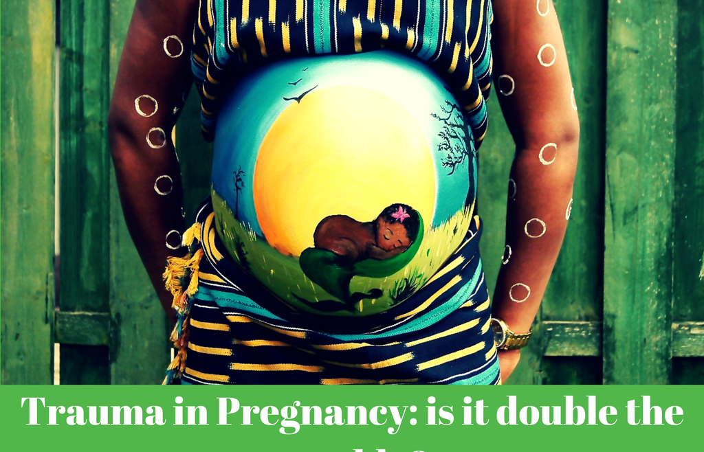
Trauma in Pregnancy: Is it double the trouble?

Managing the injured pregnant trauma patient can be anxiety inducing, as one needs to manage two patients at once amidst the altered physiology of pregnancy.  The good news is that there are more similarities than differences when managing a pregnant trauma patient. Trauma in pregnancy is surprisingly common, affecting 7% of all pregnancies. This means that if you work in an Emergency Department, you will manage an injured pregnant patient at some point. Trauma is responsible for the highest number of maternal deaths when obstetric causes are excluded. In most countries, motor vehicle accidents are the commonest type of trauma, and are also responsible for the highest number of fetal deaths. Intimate partner violence, is the second most common cause, which we should always be careful to exclude in women presenting following trauma to the ED.

So how do we look after our two patients? Is it double the trouble? It’s a big topic which will be split over three posts. First, let’s begin with resuscitation of the severely injured pregnant trauma patient.

How would you manage this case?

A young woman, in her second trimester of pregnancy, presents following an motor vehicle collision. She was not wearing a seatbelt. She arrives in the ED brought by basic life support crew, who have immobilised her on a spine board. She is diaphoretic, tachypnoiec and appears grey. She is alert but anxious, complaining of chest and abdominal pain. Initial vitals are: Sats 82% on room air, HR 120 BPM, BP 80/60.

Initial steps:

You need a multi-disciplinary team. Call for help early.

The principles of major trauma resuscitation still apply. Manage her initially as you would manage any other critically injured patient. The primary survey focuses on identifying life threatening injuries and complications and managing them.

Resuscitate the mother as your priority. All resuscitative efforts are focused on her initially, regardless of the gestational age of the fetus. Improving the mother’s oxygenation and perfusion will improve fetal oxygenation and perfusion.

The pregnant trauma patient is a physiologically and anatomically difficult airway. Prepare well and be meticulous.

Administer supplemental oxygen if required, aiming for oxygen sats of 95-98%.

Fetal loss is high when maternal shock is present. Identify the cause of the maternal shock and treat. Obtain good vascular access and activate your massive transfusion protocol early. Administer O negative blood until the mother’s Rh status is known to avoid Rhesus iso-immunisation.

The pregnant patient has an increased circulating blood volume. This means that she loses more of her blood volume to manifest hypotension than if she were not pregnant. It is easy to underestimate blood loss in a pregnant patient. Hypotension in the pregnant trauma patient is end-stage shock.

Pregnant women were excluded from trials studying the efficacy of Tranexamic acid (TXA) in trauma patients. It classified as a category B drug in pregnancy, meaning that no animal studies have shown adverse outcomes with its use, but no human studies exist. TXA crosses the placenta. Despite the lack of published research, TXA may be of benefit in bleeding, seriously injured pregnant patients.

The management of haemorrhage is to stop the haemorrhage. The indications for operative control of bleeding still apply to pregnant trauma patients as they do to non-pregnant patients. Pelvic fractures can bleed heavily due to the engorged pelvic veins. Signs of peritonism can be unreliable in pregnancy, so have a low threshold to investigate abdominal pain further.

Shock should always be considered due to haemorrhage first, but bear in mind the presence of aortocaval compression by the uterus and its effects on the mother’s haemodynamics. After 20 weeks of pregnancy, the IVC is commonly compressed by the uterus, and the aorta may be compressed in severe cases. This can cause a 20% drop in SV and Cardiac output, reducing perfusion to maternal organs, especially the placenta. This reduction in perfusion may be critical in an injured patient. Furthermore, the fetus may be hypoxic due to reduced placental perfusion even though the mother may appear normotensive. Remember to perform manual uterine displacement in all pregnant trauma patients over 20 weeks gestation.

Avoid vasopressors wherever possible. They are associated with worse outcomes in all trauma patients. Furthermore, they produce vasoconstriction of the placental bed, increasing the risk of fetal hypoxia.

Traditionally it is recommended to tilt the patient left lateral. This can be difficult to accomplish while resuscitative measures are underway.

An easier way is to keep her supine, and displace the uterus off the IVC, by either pulling the uterus from the left, or standing on the right side and pushing the uterus towards the left. Pushing from the right side is easier to do than pulling from the left, especially if it needs to be done for long periods.

The gravid uterus displaces the diaphragm upwards. Intercostal catheters should therefore be placed 1-2 intercostal spaces higher than normal to avoid inadvertent intra-abdominal placement.

Injured pregnant women should never be denied adequate analgesia. Opiates are safe in pregnancy. Anti-tetanus toxoid is also safe in pregnancy.

As soon as feasible, an obstretic ultrasound should be done to determine to determine fetal viability. The gestational age at which the fetus is considered viable varies between countries: most guidelines consider viability from 23-24 weeks. In LMICs, fetal viability may be considered to be more than 28 weeks, depending on the available resources to look after preterm infants.

If the gestational age is below than 23-24 weeks, no external fetal monitoring is advised, and further management is focused on optimising the mother. If the gestational age is more than 24 weeks, cardiotocographic monitoring is advised to monitor fetal well-being and to monitor for uterine contractions, provided that monitoring does not interfere with maternal resuscitation and care.

Resuscitative hysterotomy is indicated in cardiac arrest and should be done as soon as possible to ensure the best outcome to the mother and fetus. It is primarily done to improve return of spontaneous circulation in the mother by improving venous return and cardiac output by eliminating aortocaval compression by the gravid uterus. It also improves pulmonary mechanics by reducing pressure on the diaphragm by the abdominal contents and reduces oxygen demand. Resuscitative hysterotomy should therefore be considered from 23 weeks gestation. If there is no time to obtain an accurate gestation, feel for the height of the uterine fundus. If it is above the umbilicus, the fetus is more than 20 weeks gestation.

In summary:

  • Principles of major trauma resuscitation still apply.
  • Resuscitation and stabilisation of the mother always takes priority over fetal well-being. Fetal outcome can be optimised with good maternal resuscitation.
  • Perform manual uterine displacement in pregnant trauma patients over 20 weeks.
  • In viable pregnancies, perform external fetal monitoring.
  • Consider resuscitative hysterotomy in patients more than 23 weeks gestation.

Click on the title below to read the next post in this three part series:

Trauma in Pregnancy Part 2: Pregnancy related complications

#badEM16 – Julian Fleming – Emergency Medicine: You’re Doing It Wrong

488 558 Craig Wylie

Emergency Medicine: You’re Doing It Wrong

Julian (@EMedic6) Brings you down to reality…. REMEMBER we are in AFRICA

Can we expect the lay-person to understand what a true emergency is? If your child has a fever, it is an emergency, maybe not to the healthcare practitioner, but to that child’s mom this is the biggest emergency that she has ever faced.

We try to change the patients’ health seeking behavior… but guess what? We can’t change the patient. The question we have to ask is, are we creating our own problems with the current system that we are using?

Listen to Julian share his thoughts and ideas.

Thank you Alan Tucker from Ysterplaat Medical Supplies for sponsorship on the day

Emergency Medicine in Malawi

720 480 Jo Park-Ross

Post by Mulinda Nyirenda and Jo Park-Ross

This podcast is an interview with Dr Mulinda Nyirenda and details the current state of Emergency Medicine and pre-hospital services in Malawi, as well as the way forward and Mulinda’s hopes for the future of Malawian EM. I must admit a more than slight bias on the topic of Malawi as I grew close to the city of Blantyre where Mulinda works.

Most of the Malawian population live well below the poverty line, and are rural subsistence farmers. While the health services provided by the state are free, the hospitals often have no medicine or consumables, and these must be purchased by the patient or their family at a pharmacy. Infant and maternal mortality rates and HIV/AIDS infection rates are high, as well as a high burden of Malaria and other infectious diseases.

Thank you Mulinda for your time and for all the wonderful work you are doing for Malawi!

Emergency Medicine in Malawi

Dr. Mulinda Nyirenda is a senior consultant in the emergency department at Queen Elizabeth Central Hospital, Blantyre, Malawi. She also holds a clinical lecturer post within the University of Malawi -College of Medicine; promoting the teaching of Emergency Medicine and Acute care in the country.

Simulation as a vehicle to excellence, improved patient safety & organisational learning with David Grant

660 330 Jo Park-Ross

Dr David Grant is a simulation expert who advocates the use of simulation, including human factors and crew resource management principles, being actively used in the healthcare environment as part of a pro-active risk management system.

Traditionally, simulation doesn’t get much of a positive reception – but we actively need to change that perception of judgement. To err is human, and as such we need to ensure that our mistakes are made on mannequins, not on patients! Simulation in our work environments aims to work on the human factors – there is good evidence that poor communication in healthcare teams is associated with poor outcomes for patients (as discussed in the video).

WHY is this important?

  • Improving care: Because we need to test our systems and patient care to ensure a patient gets the very best care no matter where they are – for example when introducing a new piece of equipment such as an intra-osseous gun
  • Complexity: simulating a procedure and training as a team to react to adverse events such as equipment failures best prepares us
  • Life long learning is important for all members of the team!

Thank you to Fahmi Adams for the video!

Interested in being trained by David Grant and his team of excellent simulators?

Here’s your chance:

About David Grant

After graduating from the University of Stellenbosch, David trained in Paediatrics and Paediatric Intensive Care in South Africa, United Kingdom and Australia. He is actively involved in implementation of simulation and technology enhanced educational tools at a local, regional, national and international level. He currently works at the Bristol Royal Hospital for Children as a PICU consultant.

To learn more:

Bristol Medical Simulation Society

The International Pediatric Simulation Society

Ubuntu #4: The curious case of the ligamentum arteriosum

1063 1125 Jo Park-Ross

Author: Pierre Smit

Original presenter: Dr John Roos

Editor: Jo Park-Ross

At approximately 07H00 Ubuntu Aeromedical Service received a call on their rotor-wing platform for a young gentleman who had fallen from a height of approximately 5 meters. They were told that the incident had occurred at 06H00, and that the patient was standing on the trailer of a truck when it suddenly moved forward, causing him to fall off of the back end, head-first onto the concrete floor.

On arrival (09H55), they found the patient with typical signs of a severe traumatic brain injury:  GCS of 4/15, Cheyne-Stokes breathing, signs of a basal skull fracture, a combination of Le Forte fractures and sluggishly reactive pupils which were equal in size. However, the patient also showed signs of severe decompensated hypovolaemic shock with a low blood pressure (63/51), pale, mottled, cold and clammy skin, and a delayed capillary refill time.

One would have expected to see a ‘Cushing’s response’ to the raised intracranial pressure consequent upon the severe traumatic brain injury (a well-perfused patient with hypertension and bradycardia).   So, what happened here – how does one explain this paradoxical clinical presentation?

Shortly after the assessment of the patient was completed, the decision was made to intubate using rapid sequence intubation. However, during preparation for intubation, the patient deteriorated rapidly, went into cardiac arrest.   After resuscitation attempts proved futile, the patient was declared dead on scene. The treating paramedic then decided to consult with the Continuous Quality Improvement department at Ubuntu Aeromedical Service, to gain  clarity about the conflicting clinical signs in this mystifying “isolated” traumatic brain injury [about this extraordinarily complicated case].

An autopsy was later performed on the patient which revealed the following: confirmed base of skull fracture, intracerebral haemorrhage, pulmonary and cardiac contusions and massive bleeding within the thoracic cavity. Together, these post-mortem diagnoses paint a pretty grim picture. So the prevailing question is: what on earth happened in the chest cavity to cause such a catastrophic bleed? The answer might be a little surprising, as all that bleeding might have resulted from the traumatic failure of a single ligament, the ligamentum arteriosum.

During our foetal development the ductus arteriosus, which links the distal distal aortic arch to the pulmonary artery, is a conduit to shunt blood away from the pulmonary circulation into the systemic circulation. While in utero, oxygenation does not occur in the foetal lungs. But rather, oxygenated blood is delivered to the foetus from the mother, through the placenta and umbilical cord, before being introduced into the inferior vena cava. The ductus arteriosus serves a vital function, as perfusing the lungs when they are not being oxygenated would be fairly pointless.

After birth, the ductus arteriosus begins to close as soon as the neonate breaths spontaneously and blood is once again reintroduced into the pulmonary circulation. Over time, the ductus arteriosus undergoes a transformation from endothelial tissue to fibrous tissue, at which point it becomes known as the ligamentum arteriosum. The ligamentum arteriosum is a fixed structure which forms a ‘tether’ between the aortic isthmus and pulmonary artery.

The distal part of the aorta (as it becomes the thoracic and abdominal aorta) is a relatively mobile and ‘heavy’ structure as it is filled with a large amount of blood. Similarly, the proximal aorta behaves much in the same way, in that it is relatively mobile in comparison to the aortic arch, where it is secured by the ligamentum arteriosum.

During rapid acceleration/deceleration injuries such as motor vehicle accidents or falls from height, the mobile sites of the aorta begin to move in response to the injury, whilst the relatively fixed (or immobile) ligamentum arteriosum prevents the aortic arch from moving. This causes a tremendous shearing force on the aortic wall, and can lead to severe damage of the aortic lining as the ligamentum arteriosum tears out of the aortic wall – and in severe cases results in aortic wall rupture. A rupture of the aorta will lead to massive bleeding within the thoracic cavity, much like was seen in the patient discussed above.

A few numbers

Motor vehicle accidents and falls from height are the two most common causes for traumatic aortic rupture, followed by pedestrian vehicle accidents and crush injuries. Around 0.5% – 2% of all non-lethal motor vehicle collisions and 10% to 20% of all high-speed deceleration fatalities cause traumatic aortic rupture. When you consider these figures, the diagnosis of traumatic aortic rupture carries a considerable risk of mortality, and is immediately lethal in 80% – 90% of cases. However, in contrast to these morbid figures, if aortic rupture was detected in time and the patient was taken to a suitable hospital with sufficient surgical capabilities, the survival rate following definitive care is 60% – 80%. Therefore, prompt recognition and definitive treatment of these injuries are paramount for survival.

Identification and Diagnosis

So now to the nitty gritty, how do we identify and diagnose traumatic aortic rupture? Unfortunately diagnosis would be close to impossible in the field, with signs and symptoms too unspecific to indicate traumatic aortic rupture, apart from severe hypovolemic shock. However, practitioners in the field should keep a high index of suspicion in cases where rapid acceleration/deceleration has occurred. Radiological investigations seem to be the most useful at diagnosing traumatic aortic rupture.

Chest X-rays offer limited diagnostic value, as they can often appear close to or completely normal in traumatic aortic rupture. Evaluation for mediastinal haematoma, and by inference a major vascular injury, is the main goal of initial chest radiograph. Mediastinal widening greater than 8cm and/or 25% of the width of the thorax is the most frequent observation. However, it may not always be the most sensitive finding. More discriminating findings include any abnormality of the transverse aortic arch or loss of the aortopulmonary window. Therefore, even in those cases where the mediastinum is not widened, obscuration of the lung interface with the transverse or descending thoracic aorta should still be viewed with suspicion.

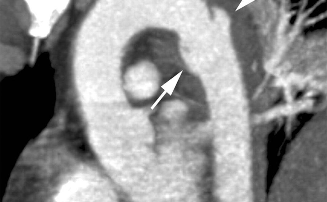
Computerised Tomography (or CT) scans are extremely useful for identifying and diagnosing traumatic aortic rupture as their contrasts between solid (tissues), fluid and gases are much clearer than chest X-rays. They offer a clear view of the continuity of the intima in the aorta, and ruptures or tears or easily visible.

Conclusion

When one considers the complexity of this case, where a severe traumatic brain injury presented together with catastrophic hypovolemic shock, it is understandable that the patient’s prognosis was very poor from the outset. However, the initial delay of an hour in activating the rotor-wing aircraft and crew, together with delays in arriving at the scene, meant that the patient had a three hour window where definitive care could have been potentially life-saving. As mentioned above, a high index of suspicion, early identification and definitive treatment are key factors to improve survival rates, and although these injuries can be immediately fatal in many instances, the survivors have a good chance of recovery if given prompt definitive care.

Pierre Smit is a flight paramedic in Cape Town South Africa who has recently completed his MPhil: Emergency Medicine at the University of Cape Town.

In his own words: “I am a passionate, fun-loving person with a desire to be as involved in Emergency Medicine education as I possibly can. I have high hopes for developing critical care in South Africa for the pre-hospital setting in the coming years, which is a goal I strive towards every day.  I am also an avid Mountain-biker and adventure-videographer with the old fractures and scars to prove it.”

Interview with the author: Hendry Sawe on recent AfJEM publication on POCUS in Tanzania & Mexico!

1900 500 Kat Evans

Series: “Interview with the Author…”

logo2

The badEM crew interviewed Hendry Robert-Sawe regarding his newly released article in AfJEM Volume 6 Issue 3 entitled: “Bedside ultrasound training at Muhimbili National Hospital in Dar es Salaam, Tanzania and Hospital San Carlos in Chiapas, Mexico” by Teri A Reynolds, Jeanne Noble, Gehres Paschal, Hendry Robert Sawe, Aparajita Sohoni, Sachita Shah, Bret Nicks, Victor Mwafongo, John Stein

Link to open access article: Click here

Corresponding author email: hendry_sawe@yahoo.com


1. Tell us about yourself. How did you get involved in this field/this research?

I have broad experience in emergency medicine & global health policy, with specific training in qualitative & quantitative research methods & a focus on emergency care system development in sub-Saharan Africa. As PI or co-investigator on university- & foundation-funded grants, I have worked to build emergency care research & clinical capacity in low & middle income countries, including the creation of open-access curricula & research instruments. I previously directed the Emergency Medicine Residency & research programs at Muhimbili National Hospital in Tanzania. As part of this project, I successfully led a multi-university-collaboration, creating the infrastructure for both educational & research initiatives that have been sustained & expanded since handover to local leadership. As Chair of the African Federation for Emergency Medicine (AFEM) Scientific Committee, I served as Editor of the AFEM Emergency Care curriculum & directed the AFEM regional Trauma Data Project. In my current role leading the emergency & trauma care program in the Department for the Management of NCDs, Disability, Violence & Injury Prevention at WHO Headquarters in Geneva, I lead a range of initiatives to assess & strengthen emergency care systems. My activities include conducting research to establish an evidence base for the impact of emergency care & to evaluate the impact of a range of system development initiatives.

2. What were the key findings from this study?

Introducing bedside ultrasound in two distinct resource-limited settings was feasible & well-received. Providers were very interested in receiving training in bedside ultrasound & completed all course sessions, even though participation significantly increased the length of their work day. After a brief intensive period of training, participants successfully passed a comprehensive examination, including demonstration of standardized image acquisition & accurate interpretation of normal & abnormal studies. Overall, participants were satisfied with the course, although there approximately one-third of providers at both sites would have preferred more hands-on training. Obstetrical, hepatobiliary & trauma applications were identified as the most useful.

3. What do these findings mean within the African context?

Ultrasound is both feasible & an affordable bedside diagnostic tool that can be deployed with minimum training to providers across Africa with significant impact to a substantial number of patients. Hospitals & programs should advocate for more use & access to bedside ultrasound availability to support care, regardless of the level of hospital (Tertiary vs Lower).

Check out the full-text open access article:  Click here


More about AfJEM (excerpt from their newsletter)

AFEMAfJEM is an open access publication in the spirit of bringing #FOAMed to Africa. This is an important consideration, especially in a low to middle income setting where prospective readers, that may benefit from published information, will most likely not be able to access subscription based journal content.  The AfJEM has no front end (author) or back end (reader) fees, & on top of that it offers a free Author Assist service that has been shown to reverse one in every four reject decisions (of manuscripts that fall within the journal’s scope) over the last five years.


More from AFEM:

Supadel (clear)
Support a delegate (Supadel) is a conference sponsorship program with a difference. Supadel is a peer-to-peer sponsorship scheme that enables prospective AfCEM2016 delegates from developed regions to financially support their peers from low & middle income countries.
For more information, to donate or to apply visit the: Sponsorship application page